Please use this identifier to cite or link to this item:
http://hdl.handle.net/10637/15264Intra-osseous plasma rich in growth factors enhances cartilage and subchondral bone regeneration in rabbits with acute full thickness chondral defects: histological assessment
| Title: | Intra-osseous plasma rich in growth factors enhances cartilage and subchondral bone regeneration in rabbits with acute full thickness chondral defects: histological assessment |
| Authors : | Torres Torrillas, Marta Damiá Giménez, Elena Romero Martínez, Ayla del Peláez Gorrea, Pau Miguel Pastor, Laura Chicharro Alcántara, Deborah Carrillo Poveda, José María. Rubio Zaragoza, Mónica. Sopena Juncosa, Joaquín Jesús. |
| Keywords: | Terapia; Therapy; Plasma sanguíneo; Blood plasma; Factores de crecimiento; Growth factors; Osteoartritis; Osteoarthritis; Cartílagos; Cartilage; Histología veterinaria; Veterinary histology |
| Publisher: | Frontiers Media |
| Citation: | Torres-Torrillas, M., Damia, E., Del Romero, A., Pelaez, P., Miguel-Pastor, L., Chicharro, D., Carrillo, J.M., Rubio, M. & Sopena, J.J. (2023). Intra-osseous plasma rich in growth factors enhances cartilage and subchondral bone regeneration in rabbits with acute full thickness chondral defects: histological assessment. Frontiers in Veterinary Science, vol. 10, art. 1131666 (23 mar.). DOI: https://doi.org/10.3389/fvets.2023.1131666 |
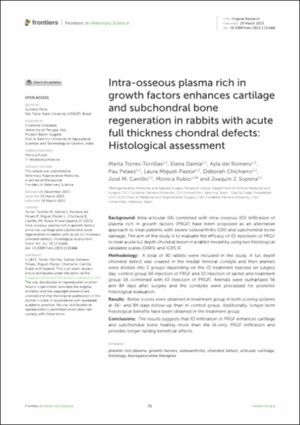
| Abstract: | Background: Intra-articular (IA) combined with intra-osseous (IO) infiltration of plasma rich in growth factors (PRGF) have been proposed as an alternative approach to treat patients with severe osteoarthritis (OA) and subchondral bone damage. The aim of the study is to evaluate the efficacy of IO injections of PRGF to treat acute full depth chondral lesion in a rabbit model by using two histological validated scales (OARSI and ICRS II). Methodology: A total of 40 rabbits were included in the study. A full depth chondral defect was created in the medial femoral condyle and then animals were divided into 2 groups depending on the IO treatment injected on surgery day: control group (IA injection of PRGF and IO injection of saline) and treatment group (IA combined with IO injection of PRGF). Animals were euthanized 56 and 84 days after surgery and the condyles were processed for posterior histological evaluation. Results: Better scores were obtained in treatment group in both scoring systems at 56- and 84-days follow-up than in control group. Additionally, longer-term histological benefits have been obtained in the treatment group. Conclusions: The results suggests that IO infiltration of PRGF enhances cartilage and subchondral bone healing more than the IA-only PRGF infiltration and provides longer-lasting beneficial effects. |
| URI: | http://hdl.handle.net/10637/15264 |
| Rights : | Open Access http://creativecommons.org/licenses/by/4.0/deed.es |
| ISSN: | 2297-1769 (Electrónico) |
| Issue Date: | 29-Mar-2023 |
| Center : | Universidad Cardenal Herrera-CEU |
| Appears in Collections: | Dpto. Medicina y Cirugía Animal |
Items in DSpace are protected by copyright, with all rights reserved, unless otherwise indicated.


