Please use this identifier to cite or link to this item:
http://hdl.handle.net/10637/10156Oxidative stress and autophagy-related changes during retinal degeneration and development
| Title: | Oxidative stress and autophagy-related changes during retinal degeneration and development |
| Authors : | Trachsel Moncho, Laura Benlloch Navarro, María Soledad Fernández Carbonell, Ángel Ramírez Lamelas, Dolores Tania Olivar Rivas, Teresa Silvestre Castelló, María Dolores Poch Jiménez, Enric Miranda Sanz, María |
| Keywords: | Retina - Enfermedades.; Enfermedades genéticas.; Genetic disorders.; Retina - Diseases.; Retinosis pigmentaria.; Estrés oxidativo.; Oxidative stress.; Retinitis pigmentosa. |
| Publisher: | Springer Nature Publishing AG. |
| Citation: | Trachsel Moncho, L., Benlloch Navarro, S., Fernández Carbonell, Á., Ramírez Lamelas, DT., Olivar, T., Silvestre, D. et al. (2018). Oxidative stress and autophagy-related changes during retinal degeneration and development. Cell Death & Disease, vol. 9, art. 812. DOI: https://doi.org/10.1038/s41419-018-0855-8 |
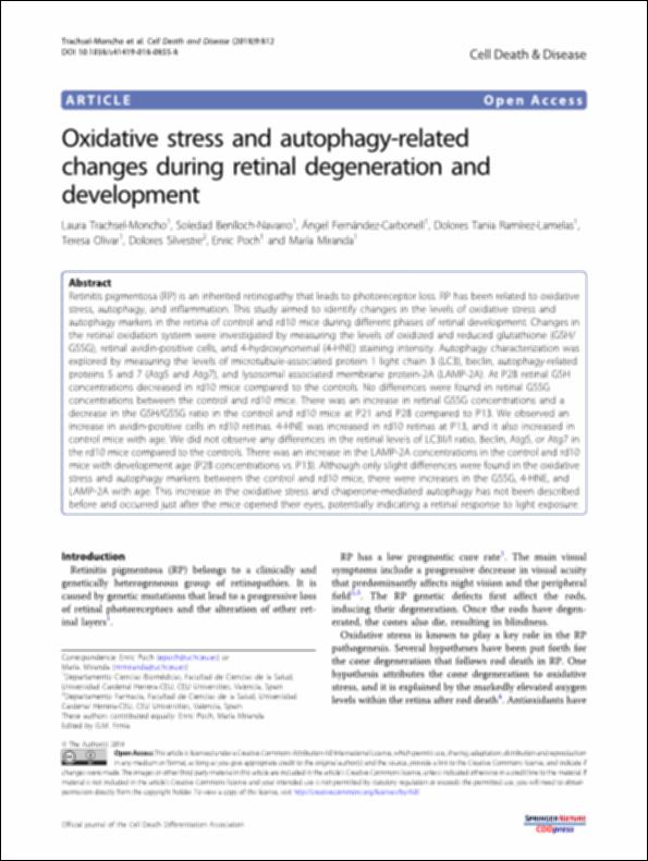
| Abstract: | Retinitis pigmentosa (RP) is an inherited retinopathy that leads to photoreceptor loss. RP has been related to oxidative stress, autophagy, and inflammation. This study aimed to identify changes in the levels of oxidative stress and autophagy markers in the retina of control and rd10 mice during different phases of retinal development. Changes in the retinal oxidation system were investigated by measuring the levels of oxidized and reduced glutathione (GSH/GSSG), retinal avidin-positive cells, and 4-hydroxynonenal (4-HNE) staining intensity. Autophagy characterization was explored by measuring the levels of microtubule-associated protein 1 light chain 3 (LC3), beclin, autophagy-related proteins 5 and 7 (Atg5 and Atg7), and lysosomal associated membrane protein-2A (LAMP-2A). At P28 retinal GSH concentrations decreased in rd10 mice compared to the controls. No differences were found in retinal GSSG concentrations between the control and rd10 mice. There was an increase in retinal GSSG concentrations and a decrease in the GSH/GSSG ratio in the control and rd10 mice at P21 and P28 compared to P13. We observed an increase in avidin-positive cells in rd10 retinas. 4-HNE was increased in rd10 retinas at P13, and it also increased in control mice with age. We did not observe any differences in the retinal levels of LC3II/I ratio, Beclin, Atg5, or Atg7 in the rd10 mice compared to the controls. There was an increase in the LAMP-2A concentrations in the control and rd10 mice with development age (P28 concentrations vs. P13). Although only slight differences were found in the oxidative stress and autophagy markers between the control and rd10 mice, there were increases in the GSSG, 4-HNE, and LAMP-2A with age. This increase in the oxidative stress and chaperone-mediated autophagy has not been described before and occurred just after the mice opened their eyes, potentially indicating a retinal response to light exposure. |
| Description: | Este artículo se encuentra disponible en la página web de la revista en la siguiente URL: https://www.nature.com/articles/s41419-018-0855-8 |
| URI: | http://hdl.handle.net/10637/10156 |
| Rights : | http://creativecommons.org/licenses/by/4.0/deed.es |
| ISSN: | 2041-4889. |
| Issue Date: | 10-Apr-2018 |
| Center : | Universidad Cardenal Herrera-CEU |
| Appears in Collections: | Dpto. Farmacia |
Items in DSpace are protected by copyright, with all rights reserved, unless otherwise indicated.


