Please use this identifier to cite or link to this item:
http://hdl.handle.net/10637/15923Morphological characterization of ckd in cats: insights of fibrogenesis to be recognized

See/Open:
Morphological_Morais_MRT_2018.jpg
561,24 kB
JPEG
See/Open:
Morphological_Morais_MRT_2018.pdf
Restricted Access
3,01 MB
Adobe PDF
Request a copy
| Title: | Morphological characterization of ckd in cats: insights of fibrogenesis to be recognized |
| Authors : | Morais, G.B. Viana, D.A. Verdugo, J.M. García Roselló, Mireia Ortega Porcel, Joaquín Rocha, D.D. Xavier Júnior, F.A.F. Barbosa, K.D.S.M Silva, F.M.O. Brito, G.A.C. Sampaio, C.M.S. Evangelista, J.S.A.M. |
| Keywords: | Enfermedad animal; Animal diseases; Riñones; Kidneys; Insuficiencia renal crónica; Chronic renal failure; Colágeno; Collagen; Gatos; Cats; Histología; Histology |
| Publisher: | John Wiley & Sons |
| Citation: | Morais, G.B., Viana, D.A., Verdugo, J.M., García Roselló, M., Ortega Porcel, J., Rocha, D.D., Xavier Júnior, F.A.F., Barbosa, K.D.S.M., Silva, F.M.O., Brito, G.A.C., Sampaio, C.M.S. & Evangelista, J.S.A.M. (2018). Morphological characterization of ckd in cats: insights of fibrogenesis to be recognized. Microscopy Research and Technique, vol. 81, i. 1 (jan.), pp. 46–57. DOI: https://doi.org/10.1002/jemt.22955 |
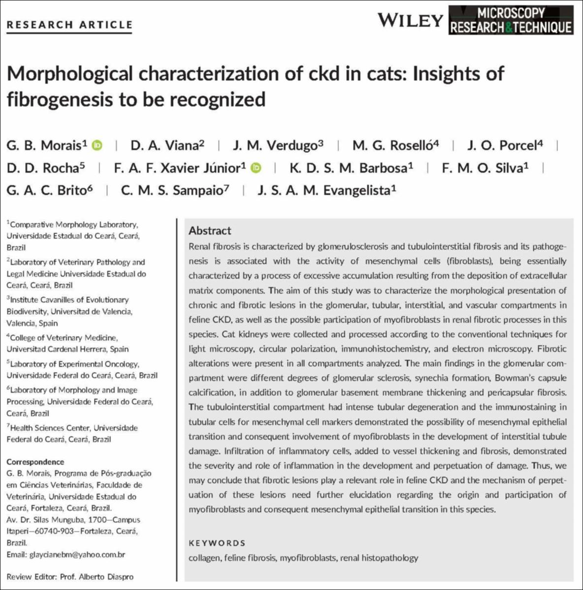
| Abstract: | Renal fibrosis is characterized by glomerulosclerosis and tubulointerstitial fibrosis and its pathoge-nesis is associated with the activity of mesenchymal cells (fibroblasts), being essentiallycharacterized by a process of excessive accumulation resulting from the deposition of extracellularmatrix components. The aim of this study was to characterize the morphological presentation ofchronic and fibrotic lesions in the glomerular, tubular, interstitial, and vascular compartments infeline CKD, as well as the possible participation of myofibroblasts in renal fibrotic processes in thisspecies. Cat kidneys were collected and processed according to the conventional techniques forlight microscopy, circular polarization, immunohistochemistry, and electron microscopy. Fibroticalterations were present in all compartments analyzed. The main findings in the glomerular com-partment were different degrees of glomerular sclerosis, synechia formation, Bowman’s capsulecalcification, in addition to glomerular basement membrane thickening and pericapsular fibrosis.The tubulointerstitial compartment had intense tubular degeneration and the immunostaining intubular cells for mesenchymal cell markers demonstrated the possibility of mesenchymal epithelialtransition and consequent involvement of myofibroblasts in the development of interstitial tubuledamage. Infiltration of inflammatory cells, added to vessel thickening and fibrosis, demonstratedthe severity and role of inflammation in the development and perpetuation of damage. Thus, wemay conclude that fibrotic lesions play a relevant role in feline CKD and the mechanism of perpet-uation of these lesions need further elucidation regarding the origin and participation ofmyofibroblasts and consequent mesenchymal epithelial transition in this species. |
| Description: | Este recurso no está disponible en acceso abierto por política de la editorial. |
| URI: | http://hdl.handle.net/10637/15923 |
| Rights : | http://creativecommons.org/licenses/by-nc-nd/4.0/deed.es |
| ISSN: | 1059-910X 1097-0029 (Electrónico) |
| Issue Date: | Jan-2018 |
| Center : | Universidad Cardenal Herrera-CEU |
| Appears in Collections: | Dpto. Medicina y Cirugía Animal |
Items in DSpace are protected by copyright, with all rights reserved, unless otherwise indicated.

