Please use this identifier to cite or link to this item:
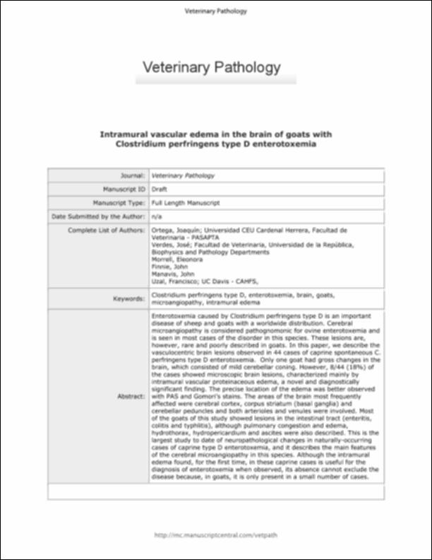
http://hdl.handle.net/10637/13870Intramural vascular edema in the brain of goats with "Clostridium perfringens" type D enterotoxemia
| Title: | Intramural vascular edema in the brain of goats with "Clostridium perfringens" type D enterotoxemia |
| Authors : | Ortega Porcel, Joaquín Verdes, José Manuel Morrell, Eleonora L. Finnie, John W. Manavis, Jim Uzal, Francisco A. |
| Keywords: | Vasos sanguíneos - Enfermedades.; Blood - Vessels - Diseases.; Cabras - Enfermedades.; Cerebro.; Clostridiosis.; Brain.; Goats - Diseases.; Clostridium diseases. |
| Publisher: | SAGE American College of Veterinary Pathologists |
| Citation: | Ortega, J., Verdes, J. M., Morrell, E. L., Finnie, J. W., Manavis, J., & Uzal, F. A. (2019). Intramural vascular edema in the brain of goats with “Clostridium perfringens” type D enterotoxemia. Veterinary Pathology, vol. 56, i. 3 (may.), pp. 452–459. DOI: https://doi.org/10.1177/0300985818817071 |
| Abstract: | Enterotoxemia caused by Clostridium perfringens type D is an important disease of sheep and goats with a worldwide distribution. Cerebral microangiopathy is considered pathognomonic for ovine enterotoxemia and is seen in most cases of the disorder in this species. These lesions are, however, rare and poorly described in goats. In this paper, we describe the vasculocentric brain lesions observed in 44 cases of caprine spontaneous C. perfringens type D enterotoxemia. Only one goat had gross changes in the brain, which consisted of mild cerebellar coning. However, 8/44 (18%) of the cases showed microscopic brain lesions, characterized mainly by intramural vascular proteinaceous edema, a novel and diagnostically significant finding. The precise location of the edema was better observed with PAS and Gomori’s stains. The areas of the brain most frequently affected were cerebral cortex, corpus striatum (basal ganglia) and cerebellar peduncles and both arterioles and venules were involved. Most of the goats of this study showed lesions in the intestinal tract (enteritis, colitis and typhlitis), although pulmonary congestion and edema, hydrothorax, hydropericardium and ascites were also described. This is the largest study to date of neuropathological changes in naturally-occurring cases of caprine type D enterotoxemia, and it describes the main features of the cerebral microangiopathy in this species. Although the intramural edema found, for the first time, in these caprine cases is useful for the diagnosis of enterotoxemia when observed, its absence cannot exclude the disease because, in goats, it is only present in a small number of cases. |
| Description: | Este artículo se encuentra disponible en la siguiente URL: https://journals.sagepub.com/doi/pdf/10.1177/0300985818817071 This is the pre-peer reviewed version of the following article: Ortega, J., Verdes, J. M., Morrell, E. L., Finnie, J. W., Manavis, J., & Uzal, F. A. (2019). Intramural vascular edema in the brain of goats with "Clostridium perfringens" Type D enterotoxemia. Veterinary Pathology, vol. 56, n. 3 (01 may.), pp. 452?459, which has been published in final form at https://doi.org/10.1177/0300985818817071 Este es el pre-print del siguiente artículo: Ortega, J., Verdes, J. M., Morrell, E. L., Finnie, J. W., Manavis, J., & Uzal, F. A. (2019). Intramural vascular edema in the brain of goats with "Clostridium perfringens" Type D enterotoxemia. Veterinary Pathology, vol. 56, n. 3 (01 may.), pp. 452?459, que se ha publicado de forma definitiva en https://doi.org/10.1177/0300985818817071 |
| URI: | http://hdl.handle.net/10637/13870 |
| Rights : | http://creativecommons.org/licenses/by-nc-nd/4.0/deed.es |
| ISSN: | 0300-9858 1544-2217 (Electrónico) |
| Issue Date: | 1-May-2019 |
| Center : | Universidad Cardenal Herrera-CEU |
| Appears in Collections: | Dpto. Producción y Sanidad Animal, Salud Pública Veterinaria y Ciencia y Tecnología de los Alimentos |
Items in DSpace are protected by copyright, with all rights reserved, unless otherwise indicated.


