Please use this identifier to cite or link to this item:
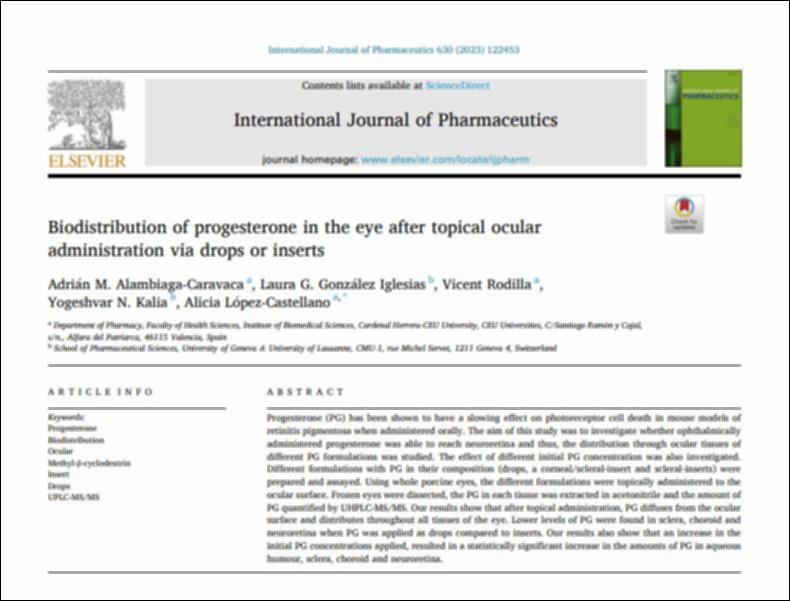
http://hdl.handle.net/10637/15067Biodistribution of progesterone in the eye after topical ocular administration via drops or inserts

See/Open:
Biodistribution_Alambiaga_IJP_2023.JPG
89,96 kB
JPEG
See/Open:
Biodistribution_Alambiaga_IJP_2023.pdf
Restricted Access
1,53 MB
Adobe PDF
Request a copy
| Title: | Biodistribution of progesterone in the eye after topical ocular administration via drops or inserts |
| Authors : | Alambiaga Caravaca, Adrián Miguel González Iglesias, Laura G. Rodilla Alama, Vicente Kalia, Yogeshvar N. López Castellano, Alicia Cristina |
| Keywords: | Medicamento; Drugs; Vista; Eyesight; Enfermedad; Diseases; Farmacología; Pharmacology |
| Publisher: | Elsevier |
| Citation: | Alambiaga-Caravaca, A.M., González Iglesias, L.G., Rodilla, V., Kalia, Y.N. & López-Castellano, A. (2023). Biodistribution of progesterone in the eye after topical ocular administration via drops or inserts. International Journal of Pharmaceutics, vol. 630, art. 122453 (05 jan.). DOI: https://doi.org/10.1016/j.ijpharm.2022.122453 |
| Abstract: | Progesterone (PG) has been shown to have a slowing effect on photoreceptor cell death in mouse models of retinitis pigmentosa when administered orally. The aim of this study was to investigate whether ophthalmically administered progesterone was able to reach neuroretina and thus, the distribution through ocular tissues of different PG formulations was studied. The effect of different initial PG concentration was also investigated. Different formulations with PG in their composition (drops, a corneal/scleral-insert and scleral-inserts) were prepared and assayed. Using whole porcine eyes, the different formulations were topically administered to the ocular surface. Frozen eyes were dissected, the PG in each tissue was extracted in acetonitrile and the amount of PG quantified by UHPLC-MS/MS. Our results show that after topical administration, PG diffuses from the ocular surface and distributes throughout all tissues of the eye. Lower levels of PG were found in sclera, choroid and neuroretina when PG was applied as drops compared to inserts. Our results also show that an increase in the initial PG concentrations applied, resulted in a statistically significant increase in the amounts of PG in aqueous humour, sclera, choroid and neuroretina. |
| Description: | Este recurso no está disponible en acceso abierto por política de la editorial. |
| URI: | http://hdl.handle.net/10637/15067 |
| Rights : | http://creativecommons.org/licenses/by-nc-nd/4.0/deed.es |
| ISSN: | 0378-5173 |
| Issue Date: | 5-Jan-2023 |
| Center : | Universidad Cardenal Herrera-CEU |
| Appears in Collections: | Dpto. Farmacia |
Items in DSpace are protected by copyright, with all rights reserved, unless otherwise indicated.

