Por favor, use este identificador para citar o enlazar este ítem:
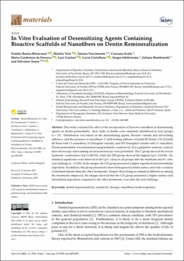
http://hdl.handle.net/10637/13573In vitro evaluation of desensitizing agents containing bioactive scaffolds of nanofibers on dentin remineralization
| Título : | In vitro evaluation of desensitizing agents containing bioactive scaffolds of nanofibers on dentin remineralization |
| Autor : | Bastos Bitencourt, Natália Velo, Marilia Nascimento, Tatiana Scotti, Cassiana Da Fonseca, Maria Gardennia Goulart, Luiz Sauro, Salvatore. |
| Materias: | Materiales nanocompuestos en Odontología.; Nanocomposites (Materials) in Dentistry.; Dentina - Enfermedades - Tratamiento.; Dentin - Diseases - Treatment. |
| Editorial : | MDPI |
| Citación : | Bastos-Bitencourt, N., Velo, M., Nascimento, T., Scotti, C., da Fonseca, M.G., Goulart, L., Castellano, L., Ishikiriama, S., Bombonatti, J. & Sauro, S. (2021). In vitro evaluation of desensitizing agents containing bioactive scaffolds of nanofibers on dentin remineralization. Materials, vol. 14, i. 5 (24 feb.), art. 1056. DOI: https://doi.org/10.3390/ma14051056 |
| Resumen : | This study evaluated the effect of the incorporation of bioactive nanofibers in desensitizing agents on dentin permeability. Sixty disks of dentin were randomly distributed in four groups (n = 15). Distribution was based on the desensitizing agents, fluoride varnish and self-etching adhesive, and the presence of nanofibers: C (self-etching adhesive Clearfil SE Bond), CN (Clearfil SE Bond with 1% nanofiber), D (Duraphat varnish), and DN (Duraphat varnish with 1% nanofiber). Dentin permeability was determined using hydraulic conductivity. For a qualitative analysis, confocal laser microscopy and scanning electron microscopy were performed. The C group showed the lowest hydraulic conductance (Lp%) (89.33), while the DN group showed the highest Lp% (116.06). No statistical significance was observed in the Lp% values in all groups after the treatment and 6% citric acid challenge (p > 0.239). In the images, the CN group presented a higher superficial and intratubular deposition. In addition, this group presented a more homogeneous dentin surface and wide occlusion of dentinal tubules than the other treatments. Despite there being no statistical differences among the treatments employed, the images showed that the CN group presented a higher surface and intratubular deposition compared to the other treatments, even after the acid challenge. |
| Descripción : | Este artículo se encuentra disponible en la siguiente URL: https://www.mdpi.com/1996-1944/14/5/1056 En este artículo de investigación también participan: Lucio Castellano, Sergio Ishikiriama y Juliana Bombonatti. Este artículo de investigación pertenece al número especial "Novel Dental Restorative Materials". |
| URI : | http://hdl.handle.net/10637/13573 |
| Derechos: | http://creativecommons.org/licenses/by/4.0/deed.es |
| ISSN : | 1996-1944 (Electrónico) |
| Fecha de publicación : | 24-feb-2021 |
| Centro : | Universidad Cardenal Herrera-CEU |
| Aparece en las colecciones: | Dpto. Odontología |
Los ítems de DSpace están protegidos por copyright, con todos los derechos reservados, a menos que se indique lo contrario.


