Por favor, use este identificador para citar o enlazar este ítem:
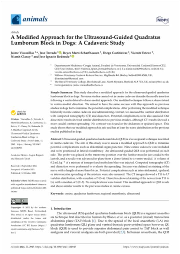
http://hdl.handle.net/10637/13395A modified approach for the ultrasound-guided quadratus lumborum block in dogs : a cadaveric study
| Título : | A modified approach for the ultrasound-guided quadratus lumborum block in dogs : a cadaveric study |
| Autor : | Viscasillas Monteagudo, Jaime Terrado Vicente, José Martí-Scharfhausen Sánchez, María de los Reyes Castiñeiras, Diego Esteve Bernet, Vicente Clancy, Niamh Redondo García, José Ignacio |
| Materias: | Anestesia local.; Anestesia veterinaria.; Veterinary anesthesia.; Quadratus lumborum muscle - Imaging.; Cuadrado lumbar - Diagnóstico por imagen.; Perros - Anestesia.; Local anesthesia.; Dogs - Anesthesia. |
| Editorial : | MDPI |
| Citación : | Viscasillas, J., Terrado, J., Marti-Scharfhausen, R., Castiñeiras, D., Esteve, V., Clancy, N., & Redondo, J. I. (2021). A modified approach for the ultrasound-guided quadratus lumborum block in dogs: a cadaveric study. Animals, vol. 11, i . 10 (12 oct), art. 2945. DOI: https://doi.org/10.3390/ani11102945 |
| Resumen : | Ultrasound-guided quadratus lumborum block (QLB) is a locoregional technique described in canine cadavers. The aim of this study was to assess a modified approach to QLB to minimise potential complications such as abdominal organ puncture. Nine canine cadavers were included and were positioned in lateral recumbency. An ultrasound-guided QLB was performed on each side. The probe was placed in the transverse position over the lumbar muscles just caudal to the last rib, and a needle was advanced in-plane from a dorso-lateral to a ventro-medial. A volume of 0.2 mL kg1 of a mixture of iomeprol and methylene blue was injected. Computed tomography (CT) and dissection were performed to evaluate the spreading. Success was defined as staining of the nerve with a length of more than 0.6 cm. Potential complications such as intra-abdominal, epidural, or intravascular spreading of the mixture were also assessed. The CT images showed a T13 to L7 vertebra distribution, with a median of 5 (3–6). Dissection showed staining of the nerves from T13 to L4, with a median of 3 (2–5). No complications were found. This modified approach to QLB is safe and shows similar results to the previous studies in canine carcass. |
| Descripción : | Este artículo se encuentra disponible en la siguiente URL: https://www.mdpi.com/2076-2615/11/10/2945 Este artículo de investigación pertenece al número que lleva por título "Loco-Regional Anaesthesia in Veterinary Medicine". |
| URI : | http://hdl.handle.net/10637/13395 |
| Derechos: | http://creativecommons.org/licenses/by/4.0/deed.es |
| ISSN : | 2076-2615 (Electrónico) |
| Fecha de publicación : | 12-oct-2021 |
| Centro : | Universidad Cardenal Herrera-CEU |
| Aparece en las colecciones: | Dpto. Medicina y Cirugía Animal |
Los ítems de DSpace están protegidos por copyright, con todos los derechos reservados, a menos que se indique lo contrario.


