Por favor, use este identificador para citar o enlazar este ítem:
http://hdl.handle.net/10637/14195Cranial thoracic myelopathies (T1-T6 vertebrae) retrospective evaluation of the signalment, clinical presentation, and, presumptive or final diagnoses in 84 dogs
| Título : | Cranial thoracic myelopathies (T1-T6 vertebrae) retrospective evaluation of the signalment, clinical presentation, and, presumptive or final diagnoses in 84 dogs |
| Autor : | Lopes, Bruno A. Ives, Edward J. José López, Roberto Gutiérrez Quintana, Rodrigo Abouzeid, Jad Freeman, Paul Redondo García, José Ignacio Sánchez Masián, Daniel |
| Materias: | Tumores en los perros - Diagnóstico por imagen.; Medula espinal - Enfermedades - Diagnóstico por imagen.; Medula espinal - Heridas y lesiones - Diagnóstico por imagen.; Spinal cord - Diseases - Imaging.; Perros - Sistema nervioso.; Dogs - Nervous system.; Spinal cord - Wounds and injuries - Imaging.; Tumors in dogs - Imaging. |
| Editorial : | Frontiers Media |
| Citación : | Lopes, B. A., Ives, E. J., José-López, R., Gutierrez-Quintana, R., Abouzeid, J., Freeman, P., Redondo, J. I. & Sánchez-Masián, D. (2022). Cranial thoracic myelopathies (T1-T6 vertebrae): retrospective evaluation of the signalment, clinical presentation, and, presumptive or final diagnoses in 84 dogs. Frontiers in Veterinary Science, vol. 9, art. 960912 (12 sep.). DOI: https://doi.org/10.3389/fvets.2022.960912 |
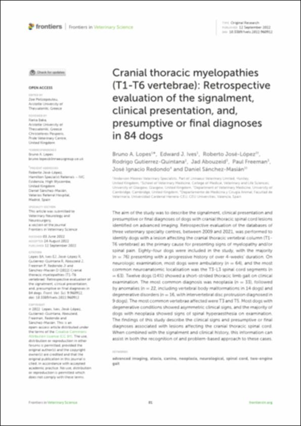
| Resumen : | The aim of the study was to describe the signalment, clinical presentation and presumptive or final diagnoses of dogs with cranial thoracic spinal cord lesions identified on advanced imaging. Retrospective evaluation of the databases of three veterinary specialty centres, between 2009 and 2021, was performed to identify dogs with a lesion aecting the cranial thoracic vertebral column (T1- T6 vertebrae) as the primary cause for presenting signs of myelopathy and/or spinal pain. Eighty-four dogs were included in the study, with the majority (n = 76) presenting with a progressive history of over 4-weeks’ duration. On neurologic examination, most dogs were ambulatory (n = 64), and the most common neuroanatomic localisation was the T3-L3 spinal cord segments (n = 63). Twelve dogs (14%) showed a short-strided thoracic limb gait on clinical examination. The most common diagnosis was neoplasia (n = 33), followed by anomalies (n = 22, including vertebral body malformations in 14 dogs) and degenerative disorders (n = 16, with intervertebral disc protrusion diagnosed in 9 dogs). Themost common vertebrae aected were T3 and T5.Most dogs with degenerative conditions showed asymmetric clinical signs, and the majority of dogs with neoplasia showed signs of spinal hyperaesthesia on examination. The findings of this study describe the clinical signs and presumptive or final diagnoses associated with lesions aecting the cranial thoracic spinal cord. When combined with the signalment and clinical history, this information can assist in both the recognition of and problem-based approach to these cases |
| Descripción : | Este artículo se encuentra disponible en la siguiente URL: https://www.frontiersin.org/articles/10.3389/fvets.2022.960912/full Este artículo pertenece a la sección "Veterinary Neurology and Neurosurgery". |
| URI : | http://hdl.handle.net/10637/14195 |
| Derechos: | http://creativecommons.org/licenses/by/4.0/deed.es |
| ISSN : | 2297-1769 (Electrónico) |
| Idioma: | es |
| Fecha de publicación : | 12-sep-2022 |
| Centro : | Universidad Cardenal Herrera-CEU |
| Aparece en las colecciones: | Dpto. Medicina y Cirugía Animal |
Los ítems de DSpace están protegidos por copyright, con todos los derechos reservados, a menos que se indique lo contrario.


