Por favor, use este identificador para citar o enlazar este ítem:
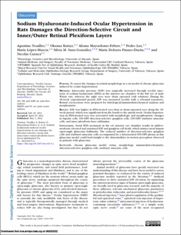
http://hdl.handle.net/10637/14167Sodium hyaluronate-induced ocular hypertension in rats damages the direction-selective circuit and inner-outer retinal plexiform layers
| Título : | Sodium hyaluronate-induced ocular hypertension in rats damages the direction-selective circuit and inner-outer retinal plexiform layers |
| Autor : | Noailles Gil, María Agustina Kutsyr, Oksana Mayordomo Febrer, Aloma Tadea Lax Zapata, Pedro López Murcia, María del Mar Sanz González, Silvia María Pinazo Durán, María Dolores Cuenca Navarro, Nicolás |
| Materias: | Eye - Diseases.; Retina - Diseases - Treatment.; Glaucoma.; Cells.; Retina.; Células.; Ojos - Enfermedades. |
| Editorial : | Association for Research in Vision and Ophthalmology (ARVO) |
| Citación : | Noailles, A., Kutsyr, O., Mayordomo-Febrer, A., Lax, P., López-Murcia, M., Sanz-González, S. M., Pinazo-Durán, M. D. & Cuenca, N. (2022). Sodium hyaluronate-induced ocular hypertension in rats damages the direction-selective circuit and inner/outer retinal plexiform layers. Investigative Ophthalmology & Visual Science, vol. 63, i. 5 (may.), art. 2. DOI: https://doi.org/10.1167/iovs.63.5.2 |
| Resumen : | PURPOSE. To assess the changes in retinal morphology in a rat model of chronic glaucoma induced by ocular hypertension. METHODS. Intraocular pressure (IOP) was surgically increased through weekly injections of sodium hyaluronate (HYA) in the anterior eye chamber of the left eye of male Wistar rats, whereas the right eyes were sham operated (salt solution). During the 10-week experimental period, IOP was measured weekly with a rebound tonometer. Retinal cryosections were prepared for histological/immunohistochemical analysis and morphometry. RESULTS. IOP was higher in HYA-treated eyes than in sham-operated eyes along the 10- week period, which was significant from the fourth to the nineth week. Ocular hypertension in HYA-treated eyes was associated with morphologic and morphometric changes in bipolar cells, ON-OFF direction-selective ganglion cells, ON/OFF starburst amacrine cells, and inner plexiform layer sublamina. CONCLUSIONS. Serial HYA treatment in the rat anterior eye chamber results in mild-tomoderate elevated and sustained IOP and ganglion cell death, which mimics most human open-angle glaucoma hallmarks. The reduced number of direction-selective ganglion cells and starburst amacrine cells accompanied by a deteriorated ON/OFF plexus in this glaucoma model could lend insight to the abnormalities in motion perception observed in patients with glaucoma. |
| Descripción : | Este artículo se encuentra disponible en la siguiente URL: https://iovs.arvojournals.org/article.aspx?articleid=2778793 |
| URI : | http://hdl.handle.net/10637/14167 |
| Derechos: | http://creativecommons.org/licenses/by-nc-nd/4.0/deed.es |
| ISSN : | 1552-5783 (Electrónico) |
| Idioma: | es |
| Fecha de publicación : | 25-may-2022 |
| Centro : | Universidad Cardenal Herrera-CEU |
| Aparece en las colecciones: | Dpto. Medicina y Cirugía Animal |
Los ítems de DSpace están protegidos por copyright, con todos los derechos reservados, a menos que se indique lo contrario.


