Por favor, use este identificador para citar o enlazar este ítem:
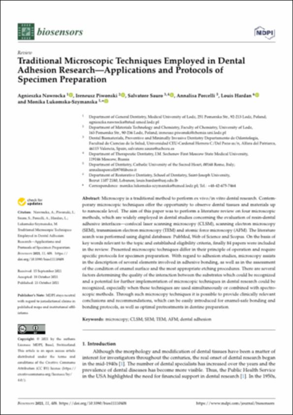
http://hdl.handle.net/10637/13570Traditional microscopic techniques employed in dental adhesion research-applications and protocols of specimen preparation
| Título : | Traditional microscopic techniques employed in dental adhesion research-applications and protocols of specimen preparation |
| Autor : | Nawrocka, Agnieszka Piwonski, Ireneusz Sauro, Salvatore. Porcelli, Annalisa Hardan, Louis Lukomska-Szymanska, Monika |
| Materias: | Adhesivos dentales - Propiedades.; Materiales dentales - Propiedades.; Microscopy.; Gums and resins, synthetic in Dentistry.; Microscopia.; Gomas y resinas sintéticas - Aplicaciones en Odontología. |
| Editorial : | MDPI |
| Citación : | Nawrocka, A., Piwonski, I., Sauro, S., Porcelli, A., Hardan, L. & Lukomska-Szymanska, M. (2021). Traditional microscopic techniques employed in dental adhesion research-applications and protocols of specimen preparation. Biosensors, vol. 11, i. 11 (21 oct.), art. 408. DOI: https://doi.org/10.3390/bios11110408 |
| Resumen : | Microscopy is a traditional method to perform ex vivo/in vitro dental research. Contemporary microscopic techniques offer the opportunity to observe dental tissues and materials up to nanoscale level. The aim of this paper was to perform a literature review on four microscopic methods, which are widely employed in dental studies concerning the evaluation of resin-dental adhesive interfaces—confocal laser scanning microscopy (CLSM), scanning electron microscopy (SEM), transmission electron microscopy (TEM) and atomic force microscopy (AFM). The literature search was performed using digital databases: PubMed, Web of Science and Scopus. On the basis of key words relevant to the topic and established eligibility criteria, finally 84 papers were included in the review. Presented microscopic techniques differ in their principle of operation and require specific protocols for specimen preparation. With regard to adhesion studies, microscopy assists in the description of several elements involved in adhesive bonding, as well as in the assessment of the condition of enamel surface and the most appropriate etching procedures. There are several factors determining the quality of the interaction between the substrates which could be recognized and a potential for further implementation of microscopic techniques in dental research could be recognized, especially when these techniques are used simultaneously or combined with spectroscopic methods. Through such microscopy techniques it is possible to provide clinically relevant conclusions and recommendations, which can be easily introduced for enamel-safe bonding and bonding protocols, as well as optimal pretreatments in dentine preparation. |
| Descripción : | Este artículo se encuentra disponible en la siguiente URL: https://www.mdpi.com/2079-6374/11/11/408 Este artículo pertenece al número especial "Multimodal, Multifunctional Biosensors or Imaging Systems and Their Biomedical Applications". |
| URI : | http://hdl.handle.net/10637/13570 |
| Derechos: | http://creativecommons.org/licenses/by/4.0/deed.es |
| ISSN : | 2079-6374 (Electrónico) |
| Fecha de publicación : | 21-oct-2021 |
| Centro : | Universidad Cardenal Herrera-CEU |
| Aparece en las colecciones: | Dpto. Odontología |
Los ítems de DSpace están protegidos por copyright, con todos los derechos reservados, a menos que se indique lo contrario.


