Por favor, use este identificador para citar o enlazar este ítem:
http://hdl.handle.net/10637/12860In vitro evaluation of antibacterial properties and smear layer removal-sealer penetration of a novel silver-citrate root canal irrigant
| Título : | In vitro evaluation of antibacterial properties and smear layer removal-sealer penetration of a novel silver-citrate root canal irrigant |
| Autor : | Tonini, Riccardo Giovarruscio, Massimo Gorni, Fabio Ionescu, Andrei Brambilla, Eugenio Mikhailovna, Irina Makeeva Luzi Luzi, Arlinda Sauro, Salvatore. |
| Materias: | Dientes - Cavidades - Preparación.; Teeth - Roots - Planing.; Terapéutica dental - Aparatos y material.; Dental therapeutics - Equipment and supplies.; Materiales dentales.; Teeth - Diseases - Treatment.; Dental materials.; Gomas y resinas - Aplicaciones en Odontología.; Gums and resins in Dentistry.; Dientes - Enfermedades - Tratamiento. |
| Editorial : | MDPI |
| Citación : | Tonini, R., Giovarruscio, M., Gorni, F., Ionescu, A., Brambilla, E., Mikhailovna, I.M. et al. (2020). In vitro evaluation of antibacterial properties and smear layer removal/sealer penetration of a novel silver-citrate root canal irrigant. Materials, vol. 13, i. 1 (02 jan.), art. 194. DOI: https://doi.org/10.3390/ma13010194 |
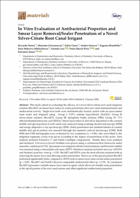
| Resumen : | This study aimed at evaluating the e cacy of a novel silver-citrate root canal irrigation solution (BioAKT) on smear layer removal, sealer penetration after root canal instrumentation and antibacterial activity. Single-root teeth were endodontically treated, sealed with an epoxi-amine resin sealer and irrigated using: Group I: 5.25% sodium hypochlorite (NaOCl); Group II: silver-citrate solution (BioAKT); Group III: phosphate bu er solution (PBS); Group IV: 17% ethylenediaminetetraacetic acid (EDTA). Smear layer removal and silver deposition at the coronal, middle and apical portion of each canal were analyzed using scanning electron microscopy (SEM) and energy-dispersive x-ray spectroscopy (EDS). Sealer penetration into dentinal tubules at coronal, middle and apical portion was assessed through dye-assisted confocal microscopy (CSM). Both SEM and CSM micrographs were evaluated by two examiners ( = 0.86), who were blind to the irrigation regimens; scores were given according to the degree of penetration of the sealer. Data analysis included Pearson’s x2 and Sidak’s multiple comparisons. Dentin discs were polished and sterilized. Enterococcus faecalis biofilms were grown using a continuous-flow bioreactor under anaerobic conditions for 72 h. Specimens were irrigated with the tested solutions, and bacterial viability was assessed using a tetrazolium salt assay (MTT). Statistical analysis included one-way ANOVA and Student’s post-hoc t-test (p < 0.05). BioAKT and EDTA were the most e cient solutions both in removing the smear layer and allowing sealer penetration. However, at the apical portion BioAKT performed significantly better compared to EDTA both in smear layer removal and sealer penetration (p < 0.05). BioAKT and NaOCl showed comparable antibacterial e ect (p = 0.53). In conclusion, BioAKT represents a suitable smear layer removal agent, which allows for reliable sealer penetration at the apical portion of the root canal system and o ers significant antibacterial properties. |
| Descripción : | Este artículo se encuentra disponible en la siguiente URL: https://www.mdpi.com/1996-1944/13/1/194 Este artículo pertenece al número especial titulado "Biomaterials for dental healing". En este artículo de investigación también participa: Paula Maciel Pires. |
| URI : | http://hdl.handle.net/10637/12860 |
| Derechos: | http://creativecommons.org/licenses/by/4.0/deed.es |
| ISSN : | 1996-1944 (Electrónico). |
| Fecha de publicación : | 2-ene-2020 |
| Centro : | Universidad Cardenal Herrera-CEU |
| Aparece en las colecciones: | Dpto. Odontología |
Los ítems de DSpace están protegidos por copyright, con todos los derechos reservados, a menos que se indique lo contrario.


